Early detection, severity grading of knee osteoarthritis using AI, novel imaging scoring method: a pilot study
A newly developed Artificial Intelligence-powered system is capable of automatically analysing MRI scans, accurately detecting the severity of knee osteoarthritis within a limited time, and even predicting potential disease progression if timely treatment is not undertaken
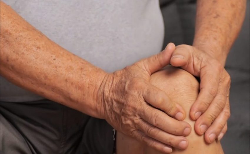
Knee pain is one of the most common complaints in Indian families, often dismissed as a natural sign of ageing or wear and tear. However, for millions of people, this pain is the first sign of knee osteoarthritis, a chronic, progressive disease that gradually damages the knee joint, making even simple tasks like walking, climbing stairs, or standing for long periods difficult. With India’s ageing population and changing lifestyle patterns marked by rising obesity and sedentary habits, knee osteoarthritis is becoming a major cause of disability and loss of independence.
One of the biggest challenges in managing this disease is the late detection. By the time most people seek medical help, the damage is already advanced, and the only remaining options are expensive and invasive, such as joint replacement surgery. Traditional methods of diagnosis rely heavily on X-rays and physical examinations, which often fail to capture early changes in the joint. Magnetic Resonance Imaging (MRI) offers a much clearer and more detailed view of the knee, particularly of the cartilage and soft tissues; however, interpreting these scans demands highly skilled radiologists, is time-consuming, and often subjective. As a result, completing and delivering timely analyses for the growing number of patients has become increasingly challenging within routine clinical schedule.
To tackle this long-standing problem, we at the Regenerative Engineering Laboratory, Indian Institute of Technology (IIT) Delhi, in collaboration with Dr Raju Vaishya at Indraprastha Apollo Hospital, New Delhi, developed an Artificial Intelligence-powered system capable of automatically analysing MRI scans, accurately detecting the severity of knee osteoarthritis within a limited time, and even predicting potential disease progression if timely treatment is not undertaken. The results of the pilot study were published recently in the journal Biomaterials Science.
Identifying, validating a new diagnostic marker
This study, conducted with full ethical approval, involved 14 patients with varying degrees of knee osteoarthritis. Each MRI scan was carefully graded by experienced radiologists using the internationally recognised MOAKS — MRI Osteoarthritis Knee Score — criteria, which assess key parameters such as cartilage thickness, bone marrow lesions (BMLs), and osteophytes (bone spurs). During this process, and with the guidance of senior radiologists from Indraprastha Apollo Hospital, we identified and validated a new and powerful parameter called “eburnation” — a polished, ivory-like hardening of the bone that occurs due to cartilage wear and direct bone-on-bone contact. While MOAKS does not traditionally emphasise eburnation as a diagnostic marker, our collaborative research with Indraprastha Apollo Hospital led to the development of an innovative scoring system capable of detecting eburnation across varying severities of osteoarthritis. Through this approach, we substantiated our hypothesis that eburnation represents the earliest and most critical sign in the progression of osteoarthritis severity.

Detecting, grading knee osteoarthritis
Using this combined approach — MOAKS criteria plus the newly discovered eburnation parameter — we trained advanced deep learning (AI) models to automatically detect and grade knee osteoarthritis. These models, inspired by how the human brain processes visual information, were trained on MRI scans from our study to recognise the patterns of disease. We tested several deep learning models, including ResNet50, DenseNet121, VGG16, and ResNet101. Among them, ResNet50 emerged as the best performer, with an accuracy of around 86% in correctly grading knee osteoarthritis into mild (grade 1), moderate (grade 2), and severe (grade 3) stages.
Compared with manual determination of the extent of damage using MRI scans, which is time consuming and is prone to human variation, the AI model quickly scans and measures cartilage thickness, identifies bone marrow lesions and osteophytes, detects early eburnation, and generates a clear severity grade. For example, in one severe case, the model detected 82% cartilage loss, 79% bone marrow lesion involvement, multiple osteophytes, and significant eburnation (eight blocks per 10 cm of bone surface). The radiologist’s manual assessment confirmed these findings, demonstrating the reliability and clinical relevance of the system. Similar accuracy was observed for patients with mild and moderate osteoarthritis, proving that the system can work across all disease stages.
This achievement holds great promise for India, where knee osteoarthritis is one of the leading causes of disability, especially in people over 45 years of age. With the number of elderly individuals rising rapidly, the burden of osteoarthritis is set to increase dramatically. Early detection can change this trajectory — lifestyle modifications, targeted exercises, and non-surgical treatments are far more effective in the early stages than in advanced disease. Our AI system can serve as a “first reader” in hospitals, screening MRI scans quickly and flagging potential cases for detailed review by doctors. This would save time for overburdened radiologists, improve the consistency of diagnosis, and ensure that patients receive timely care. What sets our project apart is its combination of international standards and novel discovery. We used the well-established MOAKS criteria as a foundation to ensure global relevance and comparability while adding the clinically validated parameter of eburnation to enhance early detection. This is not just a theoretical exercise; it is backed by ethical research and real-world hospital data.
A larger study soon
Our pilot study with 14 patients has shown strong results, but this is only the first step. We are now planning a much larger study involving 100 patients, again in collaboration with Indraprastha Apollo Hospital. In this next phase, we will not only refine the AI system for greater accuracy but also explore the changes in the parameter differences between male and female patients. There is growing evidence that osteoarthritis does not progress identically in men and women — factors such as hormonal changes, bone density, and activity patterns may influence its development. By comparing these parameters along with existing parameter across genders, we aim to develop a more personalised, data-driven approach that can guide tailored prevention and treatment strategies for each group.
The potential of this system extends beyond knees. The same AI framework could be applied to other joints commonly affected by osteoarthritis, such as the hips or spine, or even adapted for monitoring disease progression over time. Because MRI is non-invasive and does not involve harmful radiation, it can be used repeatedly to track changes without risking patient safety. Over time, as more data is collected and the system continues to learn, its accuracy will only improve. This project is a strong example of how pioneering technology, when combined with clinical expertise and ethical research practices, can directly improve people’s lives.
The ResNet50-based model we developed does not aim to replace doctors; instead, it empowers them. It enhances precision, reduces errors, and brings consistency to a process that has long been subjective and helps in saving time. While knee osteoarthritis currently has no cure, an accurate and advance detection can be the key to slowing its progression and reducing its impact. Our AI-powered MRI grading system offers a new pathway for this early detection — one that is fast, accurate, objective, and scalable — and give every patient a better chance at a pain-free, active life.

